If you’ve spent any time in mycology circles, you’ve probably heard whispers about the Jedi Mind Fuck mushroom. Known simply as JMF to those in the know, this captivating strain of Psilocybe cubensis has earned quite a reputation among microscopy enthusiasts and fungal researchers. What makes it so special? Well, that’s exactly what we’re here to explore.
Developed in the early 2000s through careful breeding, JMF stands out as a remarkable achievement in fungal genetics. For those passionate about microscopy, Jedi Mind Fuck spores provide an exceptional window into the intricate world of spore morphology and fungal biology. But before we dive deeper, let’s be crystal clear: everything we discuss here relates strictly to microscopy and taxonomy research. These spores are for scientific observation only.
Origins and History of Jedi Mind Fuck Mushroom
Every great strain has a story, and the tale of how Jedi Mind Fuck mushroom came to be reads like mycological folklore. Picture this: it’s the early 2000s, and underground mycology communities are buzzing with excitement about new strain developments. Enter our mysterious protagonist—or should we say, protagonists?
The most widely accepted origin story credits a dedicated mycologist operating under the pseudonym “Myco Joe.” Working in Georgia, USA, he reportedly stumbled upon some wild specimens that caught his trained eye. But finding interesting mushrooms is just the beginning. What followed was years of meticulous work—isolating desirable traits, stabilizing genetics, and coaxing consistency from nature’s inherent variability. Think of it as selective breeding meets scientific precision.
However, not everyone agrees on this version of events. A competing narrative has emerged from California’s mycology scene, suggesting that JMF actually arose from an intentional cross between Z-strain genetics and the ever-popular Golden Teacher mushroom. In this telling, researchers including “Myco Joe” and someone known as “John Meddler” collaborated to create this unique hybrid. Which story is true? Perhaps both contain elements of fact—such mysteries are common in the world of underground mycology.
As for the name? Well, the Star Wars reference wasn’t accidental. Early researchers noted something special about this strain that seemed to capture attention in ways other varieties didn’t. The moniker stuck, and today JMF has moved from underground curiosity to mainstream research interest, even catching the attention of FDA-cleared clinical studies in Arizona. Not bad for a mushroom with such humble beginnings.
Physical Characteristics of JMF Mushroom
Now, let’s get into what makes JMF visually distinctive. If you’re examining specimens for research purposes, knowing these physical traits is essential. Think of it as learning to spot the difference between similar-looking cousins at a family reunion—once you know what to look for, the differences become obvious.
Cap Morphology
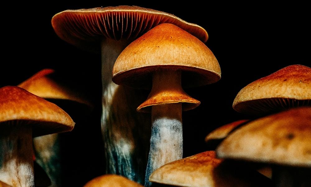
First impressions matter, and the cap is usually what catches your eye first. JMF caps sport a rich, golden-brown color that’s hard to miss. When young, they’re shaped like tiny umbrellas—technically hemispherical to convex—but here’s where it gets interesting. As they mature, these caps flatten out, almost like they’re relaxing after a long day.
Look closely at the center, and you’ll spot something distinctive: a pronounced umbo. That’s mycology-speak for a cone-shaped bump that looks a bit like a tiny hat on top of a hat. This feature becomes even more noticeable as the mushroom ages, while the cap edges start curving upward, giving mature specimens an almost whimsical appearance.
Size-wise, JMF doesn’t hold back. These caps grow notably larger than your average cubensis variety. The surface stays smooth when conditions are dry, but add a little humidity, and it develops a slightly sticky quality that photographers love to capture. Speaking of which, these photogenic features make JMF a favorite subject for research documentation.
Stem Structure
If caps are the mushroom’s crown, stems are its backbone—and JMF has quite the backbone indeed. We’re talking thick, meaty stalks that measure anywhere from 5 to 15 centimeters tall. These aren’t your delicate, wispy stems; they’re robust structures that mean business.
Color-wise, you’ll see shades ranging from pristine white to pale yellow. But here’s where things get really interesting for researchers: touch these stems (with proper laboratory gloves, of course), and you’ll witness the famous bluish bruising reaction. This color change happens when psilocybin oxidizes—a chemical calling card that helps confirm you’re dealing with authentic specimens.
Pay special attention to the stem base, where this bluing tends to be most dramatic. It’s one of those features that, once you’ve seen it, becomes an unmistakable identifier. Plus, these sturdy stems have a practical benefit: they’re perfect for creating spore prints since they won’t topple over mid-process.
Gill Formation
Flip a cap over, and you’ll discover the gills—nature’s spore factories arranged in elegant, closely-spaced lines. In young JMF specimens, these delicate structures start out whitish, almost innocent-looking. But watch them over time (a fascinating process for any researcher), and you’ll see them transform to a deep purple-brown as the spores mature.
This color progression isn’t just beautiful; it’s incredibly useful. Researchers use this visual cue to time their spore collection perfectly. Too early, and the spores aren’t ready. Too late, and you’ve missed the optimal window.
Recent studies in the Journal of Natural Products highlight how gill patterns serve as taxonomic fingerprints. In JMF’s case, the gills attach to the stem in what mycologists call an adnate to adnexed pattern—basically, they connect directly or slightly curve away from the stem. It’s a subtle detail, but these nuances separate one strain from another in the vast world of P. cubensis.
Microscopic Properties for Research
Here’s where things get truly fascinating. While the naked eye reveals impressive features, putting JMF under a microscope opens up an entirely different universe. It’s like switching from watching a movie on your phone to experiencing it in IMAX—suddenly, you’re seeing details you never knew existed.
Spore Characteristics
Let’s talk numbers for a moment. JMF spores measure between 11-17 × 8-12 micrometers—tiny measurements that place them squarely within the typical range for P. cubensis. But don’t let “typical” fool you into thinking they’re ordinary. Under magnification, these spores reveal themselves as perfect little ellipsoids, each one wrapped in thick, smooth walls that gleam under proper lighting.
What really catches the eye is their color. En masse, JMF spores create a striking dark purple to nearly black appearance that’s unmistakable once you’ve seen it. It’s nature’s own ink, concentrated and mysterious.
For those working with spore syringes, here’s a pro tip: less is more. A single drop of properly diluted spore solution on a clean slide gives you plenty to work with. Set your microscope between 400x and 1000x magnification, and prepare to be amazed. The clarity at these magnifications transforms what looks like purple-black liquid into a field of individual spores, each one a potential subject for hours of observation.
Cellular Structures
Diving deeper into the microscopic realm, we encounter the basidia—the actual spore-producing cells. In JMF, these typically appear as four-spored structures, which is exactly what we’d expect from P. cubensis. Think of them as tiny factories, each one precisely engineered to produce exactly four spores.
Then there are the cheilocystidia and pleurocystidia—admittedly tongue-twisting names for the sterile cells decorating the gill edges and faces. These structures have distinctive shapes that mycologists describe as lageniform to ventricose. In plain English? They look like tiny flasks or swollen bottles. Getting a clear view of these requires some finesse with staining techniques, but the results are worth the effort.
Modern microscopy has revolutionized how we see these structures. As the folks at Nikon MicroscopyU have documented, techniques like phase contrast and differential interference contrast microscopy work wonders with unstained JMF specimens. These methods create stunning images that reveal cellular details our mycological predecessors could only dream about.
Unique Microscopic Features
So what sets JMF apart under the lens? Several subtle but significant details emerge with careful observation. First, there’s the spore density—properly prepared JMF samples pack more spores per milliliter than many other strains. It’s like comparing a crowded concert to a half-empty theater; the difference is noticeable.
Even more intriguing are the germination patterns. Under controlled laboratory conditions, JMF spores show distinctive timing in their development stages. While we’re still gathering data on these patterns, early observations suggest they follow their own schedule, different from other cubensis varieties.
Here’s the exciting part: formal peer-reviewed studies specifically on JMF microscopy are still relatively scarce. This means citizen scientists and amateur mycologists have real opportunities to contribute meaningful observations to our collective understanding. Every carefully documented observation adds another piece to the puzzle.
Research Applications and Scientific Interest
Beyond the microscope lies a broader question: why does JMF matter to science? As it turns out, this strain offers far more than just interesting visuals. It’s become a valuable tool across multiple research disciplines, each finding unique ways to leverage its distinctive properties.
Genetic Research Potential
Think of JMF as a living laboratory for understanding fungal genetics. Because we know its backstory—the careful breeding, the selection process, the stabilization work—researchers can trace how specific traits get passed down and locked in place. It’s like having a detailed family tree that shows exactly how certain characteristics became dominant.
What makes this particularly exciting is timing. JMF emerged recently enough that we have good documentation, yet it’s been around long enough to study multiple generations. This sweet spot gives scientists a rare window into real-time evolution and adaptation.
The next frontier? DNA sequencing. Modern genetic analysis tools could pinpoint the exact markers that make JMF unique. Imagine being able to identify the specific genes responsible for those thick stems or that distinctive umbo. This isn’t just academic curiosity—understanding these genetic blueprints helps us grasp the bigger picture of how fungi evolve and diversify.
Educational Applications
Walk into any university mycology lab, and you might find students hunched over microscopes, examining JMF spores. Why this particular strain? Simple: it’s forgiving. These robust spores survive beginner mistakes that might destroy more delicate specimens. Drop your slide? Accidentally let it dry out? JMF spores can handle it.
But there’s more to it than durability. JMF’s distinctive features create those “aha!” moments that hook students on mycology. When a student successfully prepares their first slide and sees those perfect ellipsoid spores, it’s genuinely exciting. These small victories build confidence and spark curiosity.
Universities are catching on. More institutions now include diverse fungal specimens in their curriculum, moving beyond the standard teaching strains. JMF, with its clear characteristics and reliable behavior, has earned a spot in many microscopy technique courses. It’s become the teaching assistant that never calls in sick.
Comparative Studies
Science thrives on comparison, and JMF provides an excellent reference point. Researchers frequently pit it against heavy hitters like Penis Envy mushroom to map the full spectrum of P. cubensis variation. These side-by-side studies reveal fascinating patterns about how fungi respond to different selection pressures.
Consider this: JMF and Penis Envy share the same species designation, yet they look remarkably different. By studying these differences, scientists learn how flexible fungal genetics can be. It’s similar to how dog breeds, despite their vast differences, all belong to the same species. These comparisons help us understand the limits and possibilities of fungal adaptation.
The practical implications extend beyond pure science. Understanding variation within species helps predict how fungi might respond to environmental changes, develop resistance, or adapt to new conditions. Every comparison adds another data point to our growing understanding of fungal biology.
Storage and Handling Best Practices
Let’s talk about keeping your JMF spores happy and healthy. After all, even the most fascinating specimens won’t reveal their secrets if they’re not properly cared for. Good storage and handling practices aren’t just recommendations—they’re the foundation of successful research.
Optimal Storage Conditions
Your refrigerator is about to become your spores’ best friend. JMF specimens thrive in cool conditions, specifically between 2-8°C (35-46°F). That’s standard fridge temperature—no special equipment needed. Just find a quiet corner where they won’t get knocked around every time someone reaches for the milk.
Here’s a crucial tip: resist the temptation to freeze them. While it might seem logical that colder equals better preservation, freezing actually creates ice crystals that can rupture those delicate spore walls. Think of it like freezing a tomato—it might preserve it, but it’ll never be quite the same afterward.
Stored properly in the fridge, JMF spores can remain viable for months, even years. It’s remarkable how patient these microscopic subjects can be. For the full scoop on maximizing longevity, check out our detailed spore syringe storage guide. Trust us, a little care now saves a lot of disappointment later.
Handling Procedures
Time to channel your inner scientist. When working with JMF spores, cleanliness isn’t just next to godliness—it’s essential for meaningful research. Start with clean everything: slides, coverslips, tools, hands. Contamination is the enemy of good microscopy, turning what should be clear observations into guessing games.
Here’s something often overlooked: temperature acclimation. When you take those spores from the fridge, give them time to warm up to room temperature. Rushing this step leads to condensation on your slides, which is about as helpful as trying to look through a fogged-up windshield. Patience pays off with crystal-clear viewing.
The pros at the American Society for Microbiology have this down to a science. Their aseptic laboratory techniques might seem like overkill for home research, but following these guidelines elevates your work from amateur hour to legitimate science. Plus, good habits formed early stick with you.
Documentation Requirements
If it’s not documented, did it really happen? In research, your notebook becomes your memory, your proof, and sometimes your inspiration when you notice patterns you missed before. Start simple: date received, storage temperature, any environmental changes. Notice your fridge went wonky last week? Write it down.
Photography changes everything. A picture through the microscope captures details you might forget and creates a visual timeline of your observations. Modern smartphones can take surprisingly good microscope photos with a steady hand or simple adapter. These images become your visual diary, showing changes over time that written notes might miss.
Remember, you’re not just keeping records for yourself. Every observation, every note, potentially contributes to the larger understanding of JMF characteristics. Your documentation today might answer someone else’s question tomorrow. It’s citizen science at its finest—each careful observer adding their piece to the larger puzzle.
Legal Considerations for Researchers
Before we go any further, let’s have an honest conversation about the legal landscape. Understanding where you stand legally isn’t just smart—it’s absolutely essential for responsible research. The rules might seem complex at first, but once you understand the basics, navigating them becomes straightforward.
Federal Regulations
Here’s the fascinating legal quirk that makes microscopy research possible: in the United States, spores of Psilocybe cubensis, including our friend JMF, occupy a unique legal position. The spores themselves don’t contain psilocybin or psilocin—the compounds that make mature mushrooms controlled substances. It’s like having seeds for a plant that’s illegal to grow; the seeds themselves aren’t the problem.
This distinction creates a clear boundary for researchers. Observing spores under a microscope? Perfectly legal in most states. Creating spore prints for study? No problem. But the moment anyone attempts to germinate or cultivate these spores, they’ve crossed into illegal territory. Think of it as the difference between admiring a fast car in a showroom versus taking it for a joyride without permission—one’s fine, the other lands you in trouble.
The key to staying on the right side of the law is simple: stick to microscopy and taxonomy research. Period. No exceptions, no gray areas, no “what ifs.” This bright line keeps legitimate researchers safe while pursuing their scientific interests.
State-Specific Laws
Now here’s where things get a bit more complicated. While federal law creates one framework, individual states sometimes add their own rules. California, Georgia, and Idaho have decided that even spore possession crosses a line, making them no-go zones for JMF research. If you’re in one of these states, even microscopy research with these spores isn’t an option.
But wait, there’s more to consider. Laws change, sometimes quickly. What’s legal today might not be tomorrow, and vice versa. Some states have pending legislation that could shift the landscape. Others have local ordinances that add another layer of complexity. It’s like a patchwork quilt where each square has different rules.
The smart move? Before ordering any specimens, spend a few minutes checking your state’s current laws. A quick search of your state legislature’s website usually provides current information. When in doubt, err on the side of caution. No research project is worth legal complications.
International Considerations
Thinking about collaborating with researchers abroad or importing specimens from another country? Pump the brakes and do your homework first. International spore laws are all over the map—literally. What’s perfectly legal in one country might carry serious penalties in another.
Some nations treat spores just like they treat mature mushrooms—completely prohibited. Others allow them for research but require special permits or documentation. Still others have no specific regulations at all. And here’s the tricky part: even if spores are legal in both countries, import and export regulations might still apply.
The complexity here isn’t something to navigate casually. If you’re considering any international exchange of specimens, consider it like planning international travel—check the rules for both your departure and destination points. When the situation gets complicated, consulting with someone who understands international regulations isn’t overcautious; it’s smart science.
Recent Developments and Future Research
The world of JMF research isn’t standing still. In fact, recent years have brought developments that would have seemed like science fiction just a decade ago. Let’s explore what’s happening now and peek into what might be coming next.
Clinical Research Initiatives
Mark your calendars: 2024 became a watershed year for JMF research. The FDA’s clearance of a clinical study in Arizona involving JMF specimens sent ripples through the scientific community. Why does this matter? Because when the FDA gives the green light, it signals a shift from fringe interest to legitimate scientific inquiry.
The Scottsdale Research Institute isn’t just dipping their toes in the water—they’re diving in headfirst. While their focus extends beyond the microscopy work we’re discussing here, their involvement brings credibility and resources that benefit all researchers. It’s like when a famous chef visits your local farmers market; suddenly, everyone’s paying attention to what’s been there all along.
What does this mean for microscopy enthusiasts? Potentially everything. Institutional interest often translates into funding, better equipment, and more opportunities for citizen scientists to contribute meaningful data. When major research institutes take notice, the entire field benefits from the spotlight.
Technological Advances
Remember when a basic light microscope was the height of technology? Those days are long gone. Today’s researchers have access to tools that reveal JMF’s secrets in stunning detail. Confocal microscopy creates three-dimensional images that let you virtually travel through a spore. Electron microscopy pushes magnification to levels where individual molecules become visible.
But here’s what’s really exciting: these technologies aren’t just showing us more detail—they’re revealing entirely new aspects of fungal biology. Structures we couldn’t see before, behaviors we didn’t know existed, patterns that challenge our assumptions. Each technological leap forward is like getting a new pair of glasses when you didn’t even know your vision was blurry.
Advanced imaging techniques now capture time-lapse sequences showing spore germination in real-time. Fluorescent markers highlight specific cellular components, turning spores into living light shows. These aren’t just pretty pictures; they’re data-rich visualizations that answer questions we couldn’t even ask before.
Community Science Contributions
Here’s perhaps the most exciting development: the democratization of mycological research. You no longer need a university lab to make meaningful contributions. Amateur mycologists armed with decent microscopes and smartphones are documenting JMF characteristics from living rooms and garage labs around the world.
Online forums and databases have become virtual research centers where observations from Omaha can complement findings from Oslo. Someone notices an unusual spore formation, posts it online, and suddenly researchers everywhere are checking their samples. It’s collaborative science at its finest, breaking down the walls between professional and amateur researchers.
This isn’t just feel-good participation; it’s producing real results. Regional variations that professional researchers might never have discovered are being documented by local enthusiasts. Rare observations that might have been dismissed as anomalies gain credibility when multiple citizen scientists report similar findings. Every photograph, every measurement, every careful observation adds to our collective understanding.
Conclusion
What a journey we’ve taken together through the microscopic world of the Jedi Mind Fuck mushroom. From its clouded origins in underground mycology circles to its current moment in the scientific spotlight, JMF has proven itself to be far more than just a strain with a memorable name.
We’ve explored how this unique Psilocybe cubensis variety stands out from its cousins—those distinctive golden-brown caps with their pronounced umbos, the thick stems that bruise blue at a touch, and the closely-spaced gills that transform from white to purple-brown as they mature. Each feature tells a story of careful breeding and natural variation, creating a strain that’s both beautiful and scientifically significant.
Under the microscope, JMF reveals even more secrets. Those perfect ellipsoid spores, measuring 11-17 × 8-12 micrometers, offer endless hours of observation for dedicated researchers. The unique cellular structures, from four-spored basidia to flask-shaped cystidia, provide taxonomic clues that help us understand fungal diversity. And with each technological advance, from confocal to electron microscopy, we’re discovering details that previous generations of mycologists could only imagine.
But perhaps what’s most exciting about JMF isn’t what we already know—it’s what we’re still discovering. The FDA-cleared studies in Arizona, the contributions of citizen scientists worldwide, and the constant technological innovations all point to a future rich with possibility. This isn’t just academic curiosity; it’s real science happening in real-time, with opportunities for everyone from professional researchers to bedroom microscopists to contribute meaningful data.
For those ready to begin their own JMF research journey, remember the fundamentals we’ve covered. Store your specimens properly in the refrigerator (never the freezer). Handle them with clean techniques and patience. Document everything—you never know when your observation might be the one that answers a crucial question. And above all, stay within the legal boundaries: these spores are for microscopy and taxonomy research only.
The story of Jedi Mind Fuck mushroom is still being written. Every slide prepared, every photograph taken, every observation recorded adds another line to this ongoing narrative. Whether you’re drawn by scientific curiosity, fascinated by fungal biology, or simply appreciate the beauty of the microscopic world, JMF offers a gateway into understanding the incredible diversity of fungi.
As we close this guide, remember that research isn’t just about what we observe—it’s about the questions we ask and the connections we make. The Jedi Mind Fuck mushroom, with all its mysteries and revelations, reminds us that even in something as small as a spore, there’s an entire universe waiting to be explored. So grab your microscope, prepare your slides with care, and join the growing community of researchers unlocking the secrets of this remarkable strain. The force—er, the fungi—will be with you, always.